



 In this chapter: respiratory organs in animals; the human respiratory system; mechanism of breathing; respiratory volumes and capacities; exchange of gases; transport of O2 and CO2; regulation; disorders.
Respiratory organs in animals
| Animals | Mechanism |
|---|---|
| Sponges, coelenterates, flatworms | Simple diffusion over the whole body surface |
| Earthworms | Moist cuticle |
| Insects | Network of tracheal tubes |
| Most aquatic arthropods and molluscs; fishes | Gills (branchial respiration) |
| Amphibians | Lungs and moist skin (cutaneous respiration) |
| Reptiles, birds, mammals | Lungs (pulmonary respiration) |
The human respiratory system
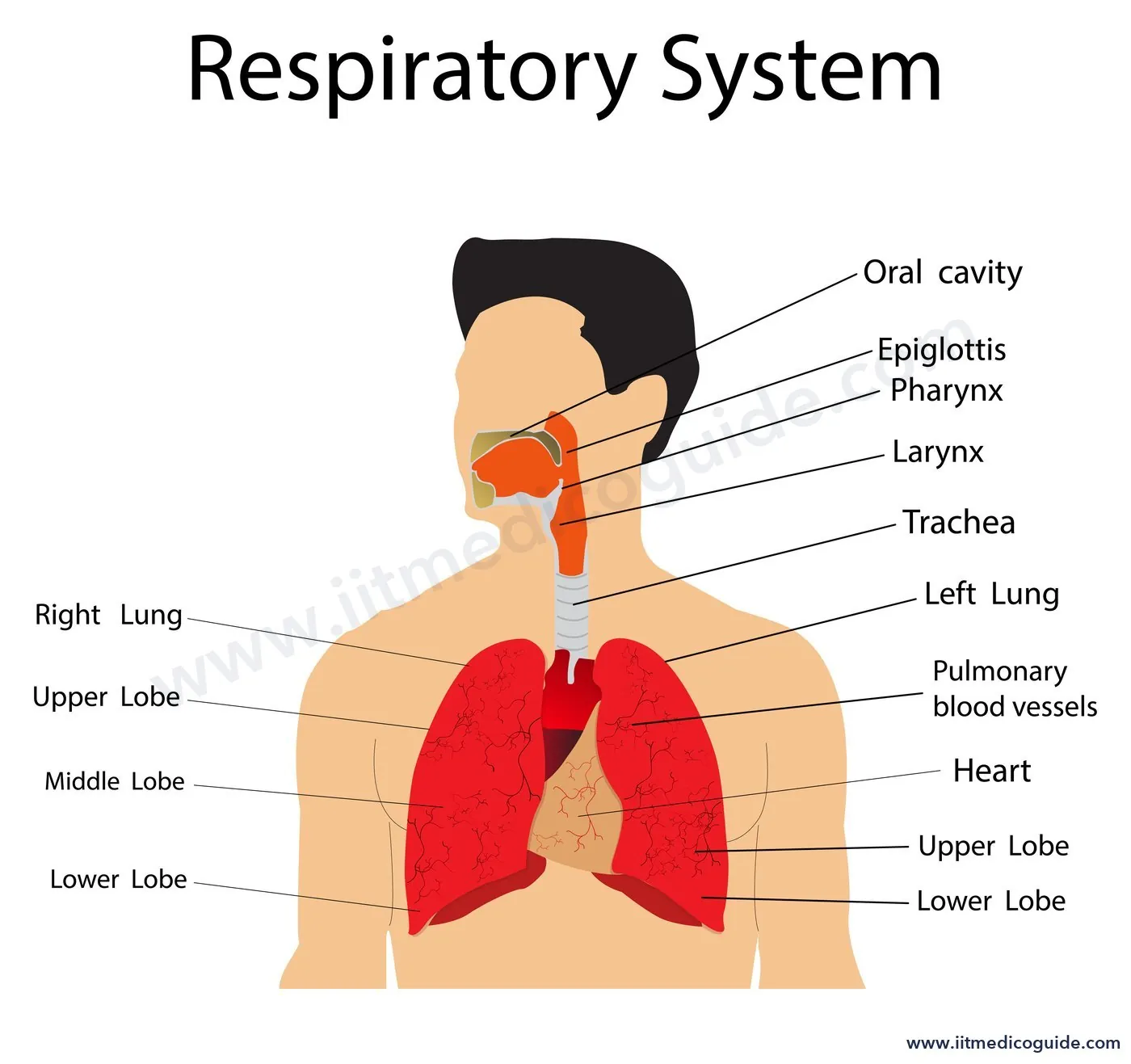
External nostrils → nasal chamber → pharynx (common passage for food and air) → larynx (cartilaginous box that produces sound: the voice box) → trachea → primary bronchi → secondary and tertiary bronchi → bronchioles → terminal bronchioles → alveoli.
- During swallowing, the glottis is covered by a thin, elastic cartilaginous flap, the epiglottis, to stop food entering the larynx.
- The trachea runs to the mid-thoracic cavity and divides at the level of the 5th thoracic vertebra into right and left primary bronchi.
- The trachea, primary, secondary and tertiary bronchi and the initial bronchioles are supported by incomplete cartilaginous rings.
- Each terminal bronchiole ends in many thin, irregular-walled, vascularised bags: the alveoli. The branching network of bronchi, bronchioles and alveoli forms the lungs.
- Lungs are covered by a double-layered pleura with pleural fluid between the layers, which reduces friction on the lung surface.
- Conducting part (nostrils to terminal bronchioles): carries air, clears it of foreign particles, humidifies it and brings it to body temperature. Respiratory (exchange) part: alveoli and their ducts, where gases diffuse.
- The lungs lie in an air-tight thoracic chamber: vertebral column behind, sternum in front, ribs on the sides and the dome-shaped diaphragm below.
Mechanism of breathing
| Inspiration | Expiration | |
|---|---|---|
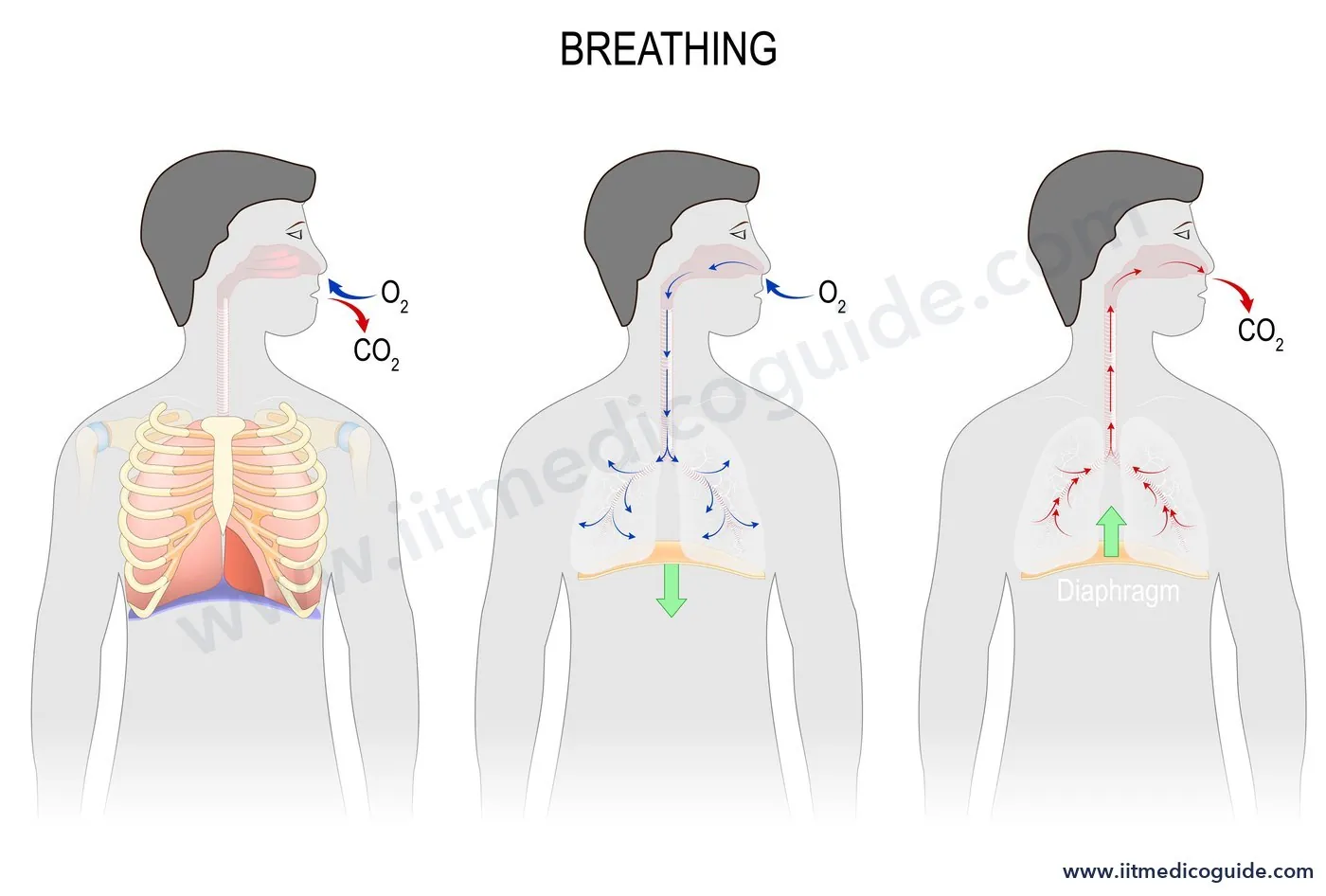
| Muscles | Diaphragm contracts (volume increases along the antero-posterior axis); external intercostal muscles contract, lifting ribs and sternum (volume increases along the dorso-ventral axis) | Diaphragm and intercostal muscles relax; thoracic volume returns to normal |
| Pressure | Intra-pulmonary pressure falls below atmospheric; air rushes in | Intra-pulmonary pressure rises above atmospheric; air is pushed out |
Additional abdominal muscles can increase the strength of both. A healthy person breathes 12 to 16 times a minute. Volumes are measured with a spirometer.
Respiratory volumes and capacities
| Term | Meaning | Value (approx.) |
|---|---|---|
| Tidal volume (TV) | Air inspired or expired in a normal breath | 500 mL (6000 to 8000 mL per minute) |
| Inspiratory reserve volume (IRV) | Additional air that can be inspired by forcible inspiration | 2500 to 3000 mL |
| Expiratory reserve volume (ERV) | Additional air that can be expired by forcible expiration | 1000 to 1100 mL |
| Residual volume (RV) | Air remaining in the lungs even after forcible expiration | 1100 to 1200 mL |
| Inspiratory capacity (IC) | TV + IRV | – |
| Expiratory capacity (EC) | TV + ERV | – |
| Functional residual capacity (FRC) | ERV + RV | – |
| Vital capacity (VC) | ERV + TV + IRV: the maximum air a person can breathe out after a forced inspiration | – |
| Total lung capacity (TLC) | RV + ERV + TV + IRV, or VC + RV | – |
 Common trap: residual volume cannot be measured by a simple spirometer, and it is not part of vital capacity. FRC includes RV; IC does not.
Exchange of gases
Gases are exchanged by simple diffusion, in the alveoli and in the tissues, driven by partial pressure gradients (pO2, pCO2). The solubility of CO2 is 20 to 25 times higher than that of O2, so much more CO2 can diffuse per unit difference in partial pressure.
| Partial pressure (mm Hg) | Atmospheric air | Alveoli | Deoxygenated blood | Oxygenated blood | Tissues |
|---|---|---|---|---|---|
| O2 | 159 | 104 | 40 | 95 | 40 |
| CO2 | 0.3 | 40 | 45 | 40 | 45 |
The diffusion membrane has three layers: the thin squamous epithelium of the alveoli, the endothelium of the alveolar capillaries, and the basement substance between them. Its total thickness is much less than a millimetre.
Transport of gases
Oxygen
- About 97% is carried by RBCs as oxyhaemoglobin; about 3% is dissolved in plasma.
- Haemoglobin is a red, iron-containing pigment; each molecule can carry at most four O2 molecules.
- Binding depends mainly on pO2, and also on pCO2, H+ concentration and temperature. Plotting % saturation against pO2 gives the sigmoid oxygen dissociation curve.
- In the alveoli (high pO2, low pCO2, fewer H+, lower temperature), oxyhaemoglobin forms. In the tissues (low pO2, high pCO2, more H+, higher temperature), it dissociates and releases O2.
- Every 100 mL of oxygenated blood delivers about 5 mL of O2 to the tissues under normal conditions.
Carbon dioxide
- About 20 to 25% is carried by haemoglobin as carbamino-haemoglobin; about 70% as bicarbonate; about 7% dissolved in plasma.
- High pCO2 and low pO2 in tissues favour CO2 binding; the reverse in the alveoli releases it.
- RBCs are rich in carbonic anhydrase (small amounts also in plasma), which speeds up: CO2 + H2O ⇌ H2CO3 ⇌ HCO3− + H+. The reaction runs forwards in tissues and backwards in the alveoli.
- Every 100 mL of deoxygenated blood delivers about 4 mL of CO2 to the alveoli.
Regulation of respiration
- The respiratory rhythm centre is in the medulla.
- The pneumotaxic centre in the pons can moderate the rhythm centre, reducing the duration of inspiration and altering the respiratory rate.
- A chemosensitive area next to the rhythm centre is highly sensitive to CO2 and H+; an increase activates the centre to remove them. Receptors in the aortic arch and carotid artery also detect changes in CO2 and H+.
- The role of oxygen in regulating the respiratory rhythm is quite insignificant.
Disorders
- Asthma: difficulty in breathing and wheezing due to inflammation of bronchi and bronchioles.
- Emphysema: a chronic disorder in which alveolar walls are damaged, reducing the respiratory surface. Cigarette smoking is a major cause.
- Occupational respiratory disorders: in industries involving grinding or stone-breaking, long exposure to dust can cause inflammation leading to fibrosis (proliferation of fibrous tissue) and serious lung damage. Workers should wear protective masks.
NEET focus
- Respiratory volumes and capacities, including which sums make which capacity.
- The partial pressure table; solubility of CO2 vs O2.
- Percentages of O2 and CO2 transport forms; 5 mL and 4 mL per 100 mL.
- Factors favouring oxyhaemoglobin formation and dissociation.
- Medulla vs pons; chemoreceptors; disorders.
Practice questions
Vital capacity equals:
- TV + IRV
- TV + ERV + RV
- ERV + TV + IRV
- ERV + RV
Show answer
The pO2 in alveoli is about:
- 159 mm Hg
- 104 mm Hg
- 95 mm Hg
- 40 mm Hg
Show answer
About 70% of CO2 is transported as:
- Carbamino-haemoglobin
- Dissolved CO2
- Bicarbonate
- Carbonic acid in RBCs
Show answer
Which condition favours dissociation of oxyhaemoglobin?
- High pO2, low pCO2
- Low temperature, fewer H+
- Low pO2, high pCO2, high H+, higher temperature
- High pO2, low temperature
Show answer
The pneumotaxic centre is located in the:
- Medulla
- Pons
- Cerebellum
- Hypothalamus
Show answer
During inspiration, the diaphragm:
- Relaxes and moves up
- Contracts and flattens, increasing antero-posterior volume
- Has no role
- Relaxes and increases pressure
Show answer
Emphysema is characterised by:
- Inflammation of bronchi causing wheezing
- Damage to alveolar walls, reducing respiratory surface
- Fibrosis due to dust
- Infection of the pleura