

 In this chapter: plasma and formed elements; ABO and Rh blood groups; coagulation; lymph; types of circulation; the human heart and its conducting system; cardiac cycle; ECG; double circulation; regulation; disorders.
Blood
Blood is a special connective tissue made of a fluid matrix (plasma) and formed elements.
Plasma (about 55% of blood)
- A straw-coloured, viscous fluid: 90 to 92% water and 6 to 8% proteins.
- Proteins: fibrinogen (clotting), globulins (defence) and albumins (osmotic balance).
- Also minerals (Na+, Ca2+, Mg2+, HCO3−, Cl−), glucose, amino acids and lipids in transit, and clotting factors in an inactive form.
- Serum = plasma without the clotting factors.
Formed elements (about 45%)
| Element | Count / share | Key facts |
|---|---|---|
| RBCs (erythrocytes) | 5 to 5.5 million per mm3 | Formed in red bone marrow in adults; no nucleus in most mammals; biconcave. Haemoglobin 12 to 16 g per 100 mL. Average life span 120 days; destroyed in the spleen (graveyard of RBCs) |
| WBCs (leucocytes) | 6000 to 8000 per mm3 | Nucleated, generally short-lived. Granulocytes and agranulocytes (below) |
| Platelets (thrombocytes) | 1.5 to 3.5 lakh per mm3 | Cell fragments of megakaryocytes in bone marrow; release clotting factors. A fall in number causes clotting disorders and excessive blood loss |
| WBC type | % of WBCs | Role |
|---|---|---|
| Neutrophils (granulocyte) | 60 to 65 (most abundant) | Phagocytic; destroy foreign organisms |
| Eosinophils (granulocyte) | 2 to 3 | Resist infections; associated with allergic reactions |
| Basophils (granulocyte) | 0.5 to 1 (least) | Secrete histamine, serotonin, heparin; involved in inflammation |
| Lymphocytes (agranulocyte) | 20 to 25 | B and T lymphocytes; immune responses |
| Monocytes (agranulocyte) | 6 to 8 | Phagocytic |
Blood groups
ABO grouping
This grouping is based on the presence or absence of two surface antigens, A and B, on RBCs. Plasma contains the opposite natural antibodies.
| Blood group | Antigens on RBC | Antibodies in plasma | Can receive from |
|---|---|---|---|
| A | A | anti-B | A, O |
| B | B | anti-A | B, O |
| AB | A, B | None | AB, A, B, O (universal recipient) |
| O | None | anti-A, anti-B | O only (O is the universal donor) |
Rh grouping
The Rh antigen is similar to one found in Rhesus monkeys. Nearly 80% of humans are Rh positive. An Rh-negative person exposed to Rh-positive blood forms antibodies against it.
Erythroblastosis foetalis can occur when an Rh-negative mother carries an Rh-positive foetus. In the first pregnancy, the mother is not exposed because the placenta keeps the two bloods separate, but during delivery she may be exposed to small amounts of foetal blood and start making anti-Rh antibodies. In later pregnancies these antibodies can leak into the foetal blood and destroy foetal RBCs, causing severe anaemia and jaundice in the baby. It is prevented by giving the mother anti-Rh antibodies immediately after the first delivery.
Coagulation of blood
- Injury stimulates platelets (and damaged tissues) to release factors that start a cascade of enzyme reactions involving many plasma factors.
- This forms the enzyme complex thrombokinase.
- Thrombokinase converts inactive prothrombin into active thrombin.
- Thrombin converts soluble fibrinogen into insoluble fibrin threads, which trap dead and damaged formed elements to form a clot (coagulum).
Calcium ions play an important role in clotting.
Lymph (tissue fluid)
As blood passes through capillaries, water and small water-soluble substances move out into the spaces between cells, leaving larger proteins and most formed elements behind. This interstitial (tissue) fluid has the same mineral distribution as plasma. All exchange of nutrients and gases between blood and cells happens through it. A network of vessels, the lymphatic system, collects it and drains it back into major veins. Lymph is colourless and contains specialised lymphocytes. It carries nutrients and hormones; fats are absorbed through lymph in the lacteals of intestinal villi.
Types of circulation
- Open: blood pumped into open spaces (sinuses): arthropods and molluscs. Closed: blood always in vessels: annelids and chordates.
- Fishes: two-chambered heart (1 atrium, 1 ventricle); single circulation (heart pumps deoxygenated blood to the gills, oxygenated blood goes to the body, deoxygenated blood returns).
- Amphibians and reptiles (except crocodiles): three-chambered heart (2 atria, 1 ventricle); oxygenated and deoxygenated blood mix in the ventricle: incomplete double circulation.
- Crocodiles, birds and mammals: four-chambered heart; oxygenated and deoxygenated blood are fully separated: double circulation.
The human heart
- Mesodermal in origin; in the thoracic cavity between the lungs, slightly tilted to the left; about the size of a clenched fist.
- Protected by a double-walled pericardium enclosing pericardial fluid.
- Four chambers: two small upper atria and two larger lower ventricles. A thin inter-atrial septum separates the atria; a thick inter-ventricular septum separates the ventricles; a thick fibrous atrio-ventricular septum separates the atrium and ventricle of each side.
- Valves: tricuspid (three cusps, between right atrium and right ventricle); bicuspid or mitral (between left atrium and left ventricle); semilunar valves at the openings of the right ventricle into the pulmonary artery and the left ventricle into the aorta. Valves allow flow in one direction only.
The conducting system
- Sino-atrial node (SAN): in the right upper corner of the right atrium.
- Atrio-ventricular node (AVN): in the lower left corner of the right atrium, close to the atrio-ventricular septum.
- The AV bundle (bundle of His) runs from the AVN through the AV septum to the top of the inter-ventricular septum, divides into right and left bundle branches, which give rise to Purkinje fibres throughout the ventricular muscle.
- Nodal tissue is autoexcitable. The SAN generates the maximum number of action potentials, 70 to 75 per minute, so it is the pacemaker. The normal heart rate is about 72 beats per minute.
The cardiac cycle
- Joint diastole: all four chambers relaxed. Tricuspid and bicuspid valves are open; blood flows from the pulmonary veins and venae cavae through the atria into the ventricles. Semilunar valves are closed.
- Atrial systole: the SAN fires; the atria contract, increasing the flow into the ventricles by about 30%.
- Ventricular systole: the impulse passes via the AVN and AV bundle to the ventricles, which contract; the atria relax at the same time. Rising ventricular pressure closes the AV valves (first sound, "lub") and then opens the semilunar valves, pushing blood into the pulmonary artery and aorta.
- Ventricular diastole: ventricles relax; pressure falls, the semilunar valves close (second sound, "dub"), then the AV valves open and the cycle repeats.
 One cardiac cycle takes 0.8 s. Stroke volume (blood pumped by each ventricle per beat) is about 70 mL. Cardiac output = stroke volume × heart rate ≈ 70 × 72 ≈ 5000 mL (5 L) per minute.
Electrocardiogram (ECG)
| Wave | Represents |
|---|---|
| P wave | Electrical excitation (depolarisation) of the atria, leading to atrial contraction |
| QRS complex | Depolarisation of the ventricles, initiating ventricular contraction. Contraction starts shortly after Q and marks the beginning of systole |
| T wave | Return of the ventricles from the excited to the normal state (repolarisation). The end of T marks the end of systole |
Counting QRS complexes over a given time gives the heart rate. Deviations from the normal shape indicate abnormalities, so the ECG is clinically important.
Double circulation and special systems
- Pulmonary circulation: right ventricle → pulmonary artery → lungs → pulmonary veins → left atrium.
- Systemic circulation: left ventricle → aorta → arteries, arterioles, capillaries → tissues → veins → venae cavae → right atrium.
- Hepatic portal system: the hepatic portal vein carries blood from the intestine to the liver before it reaches the systemic circulation.
- Coronary system: vessels that serve the heart muscle itself.
Regulation of cardiac activity
Heart activity is auto-regulated by the nodal tissue: the heart is myogenic. A cardiovascular centre in the medulla oblongata can moderate it through the autonomic nervous system: sympathetic nerves increase the heart rate, the strength of ventricular contraction and the cardiac output; parasympathetic signals decrease the heart rate and the speed of conduction, reducing cardiac output. Adrenal medullary hormones can also increase cardiac output.
Disorders
- Hypertension: blood pressure higher than normal (120/80 mm Hg). Repeated readings of 140/90 or higher indicate hypertension, which can lead to heart disease and damage vital organs like the brain and kidneys.
- Coronary artery disease (atherosclerosis): deposits of calcium, fat, cholesterol and fibrous tissue narrow the vessels supplying the heart muscle.
- Angina (angina pectoris): acute chest pain when not enough oxygen reaches the heart muscle; more common in middle-aged and elderly people.
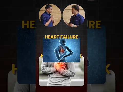
- Heart failure: the heart does not pump blood effectively enough; also called congestive heart failure because lung congestion is a main symptom. It is not the same as cardiac arrest (the heart stops beating) or a heart attack (heart muscle suddenly damaged by inadequate blood supply).
 Common traps: (1) "Lub" = closure of AV valves; "dub" = closure of semilunar valves. (2) AVN is in the right atrium. (3) Neutrophils are the most abundant WBCs; basophils the least. (4) Serum lacks clotting factors, not all proteins. (5) Crocodiles have a four-chambered heart.
NEET focus
- Plasma proteins and their roles; counts and life span of formed elements; WBC percentages.
- ABO table; Rh incompatibility and its prevention.
- Clotting cascade order and the role of Ca2+.
- Location of SAN and AVN; cardiac cycle events; cardiac output calculation; ECG waves.
- Types of circulation across vertebrates; hepatic portal system; disorders.
Practice questions
The most abundant WBCs are:
- Lymphocytes
- Neutrophils
- Monocytes
- Eosinophils
Show answer
A person with blood group AB has:
- Both anti-A and anti-B antibodies
- No antibodies in plasma
- Only anti-A
- No antigens
Show answer
The QRS complex of an ECG represents:
- Atrial depolarisation
- Ventricular depolarisation
- Ventricular repolarisation
- Atrial repolarisation
Show answer
If the stroke volume is 70 mL and heart rate 72 per minute, the cardiac output is about:
- 1 L/min
- 5 L/min
- 10 L/min
- 0.8 L/min
Show answer
The second heart sound "dub" is due to closure of:
- Tricuspid valve
- Bicuspid valve
- Semilunar valves
- AV valves
Show answer
Thrombin converts:
- Prothrombin to thrombin
- Fibrinogen to fibrin
- Fibrin to fibrinogen
- Thrombokinase to thrombin
Show answer
Incomplete double circulation is seen in:
- Fishes
- Frogs
- Crocodiles
- Birds