

 In this chapter: male and female reproductive systems; spermatogenesis and its hormonal control; structure of sperm; oogenesis; menstrual cycle; fertilisation and sex determination; cleavage and implantation; placenta and pregnancy; parturition and lactation.
The male reproductive system
- Testes lie outside the abdomen in the scrotum, which keeps them 2 to 2.5 °C below body temperature, necessary for spermatogenesis. An adult testis is oval, 4 to 5 cm long and 2 to 3 cm wide.
- Each testis has about 250 testicular lobules, each with 1 to 3 highly coiled seminiferous tubules. The tubules are lined by spermatogonia (male germ cells) and Sertoli cells (which nourish the germ cells).
- Between the tubules, the interstitial spaces contain blood vessels and Leydig (interstitial) cells, which make androgens, plus some immunologically competent cells.
- Duct path: seminiferous tubules → rete testis → vasa efferentia → epididymis (on the posterior surface of the testis) → vas deferens (loops over the urinary bladder, receives the duct of the seminal vesicle) → ejaculatory duct → urethra → urethral meatus.
- The penis has special tissue for erection; its enlarged end, the glans penis, is covered by the foreskin.
- Accessory glands: paired seminal vesicles, one prostate and paired bulbourethral glands. Their secretions form the seminal plasma, rich in fructose, calcium and certain enzymes. Bulbourethral secretions also lubricate the penis.
The female reproductive system
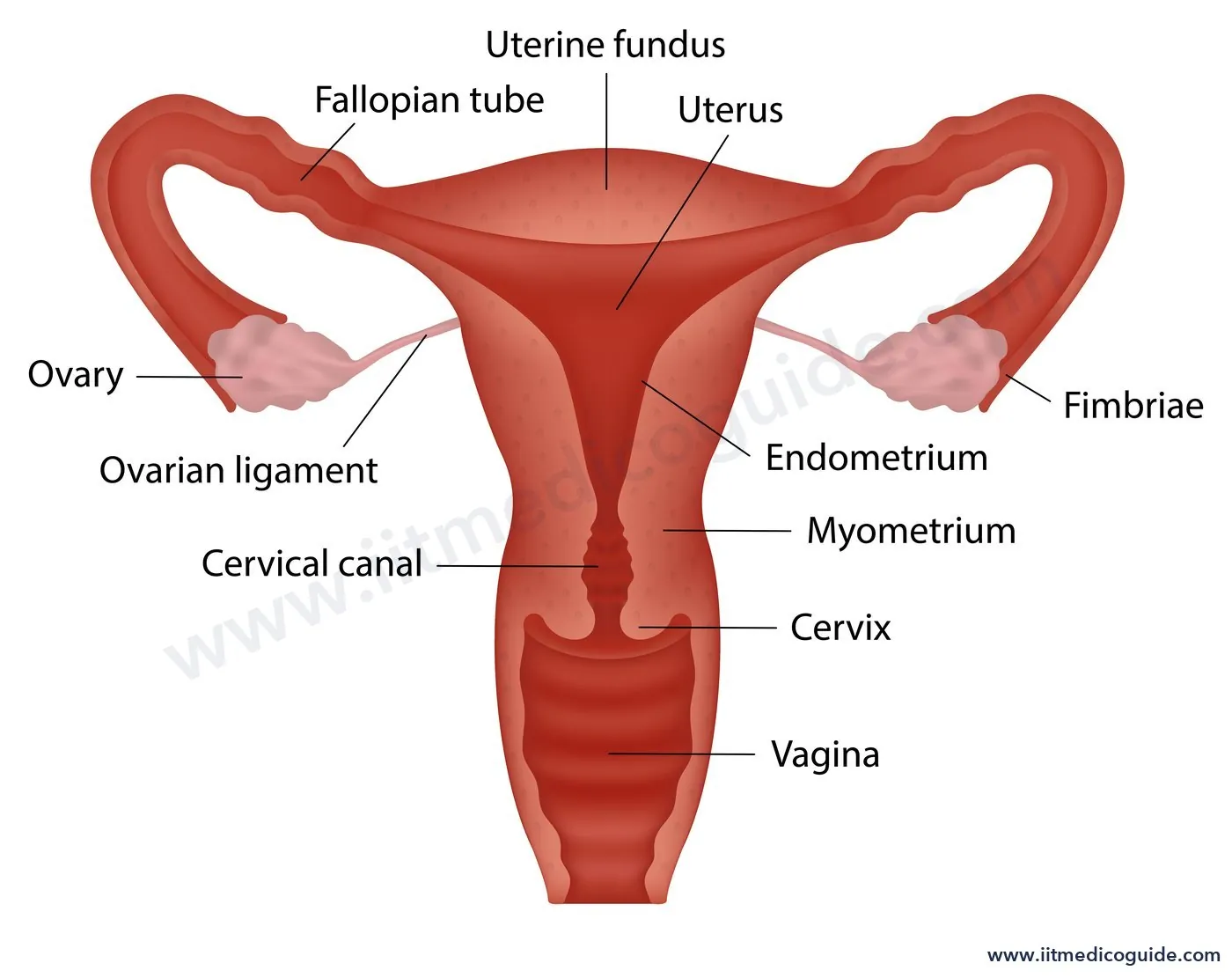
- Ovaries: the primary female sex organs, one on each side of the lower abdomen, 2 to 4 cm long, attached to the pelvic wall and uterus by ligaments. Covered by thin epithelium enclosing the ovarian stroma (outer cortex, inner medulla).
- Oviducts (fallopian tubes): 10 to 12 cm long. From the ovary end: the funnel-shaped infundibulum with finger-like fimbriae (which collect the ovum after ovulation), the wider ampulla, and the narrow isthmus joining the uterus.
- Uterus (womb): single, shaped like an inverted pear. It opens into the vagina through the narrow cervix; the cervical canal and vagina form the birth canal. The uterine wall has three layers: outer perimetrium, middle myometrium (thick smooth muscle that contracts strongly during delivery) and inner glandular endometrium (which changes cyclically during the menstrual cycle).
- External genitalia: mons pubis, labia majora, labia minora, hymen and clitoris. The hymen may be torn during first coitus but also by a fall, jolt, tampon use or sports like cycling or horse-riding, and in some women it persists after coitus, so it is not a reliable indicator of virginity.
- Mammary glands: glandular tissue divided into 15 to 20 mammary lobes containing clusters of alveoli, which secrete milk. Path of milk: alveoli → mammary tubules → mammary duct → mammary ampulla → lactiferous duct.
Spermatogenesis
- Begins at puberty. Spermatogonia (2n = 46) on the inner wall of seminiferous tubules multiply by mitosis.
- Some become primary spermatocytes (2n = 46), which complete meiosis I to form two equal, haploid secondary spermatocytes (n = 23).
- Meiosis II gives four equal, haploid spermatids.
- Spermatids are transformed into spermatozoa by spermiogenesis. Their heads stay embedded in Sertoli cells until they are released from the tubules: spermiation.
Hormonal control
At puberty, GnRH from the hypothalamus increases and acts on the anterior pituitary to release LH and FSH. LH acts on Leydig cells to secrete androgens, which stimulate spermatogenesis. FSH acts on Sertoli cells to secrete factors that help spermiogenesis.
The sperm
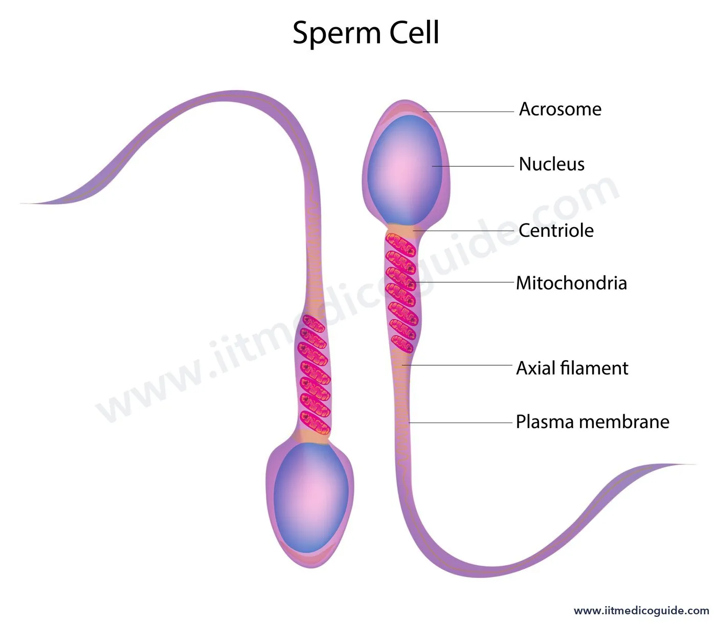
- A sperm has a head, neck, middle piece and tail, all covered by the plasma membrane.
- The head has an elongated haploid nucleus capped by the acrosome, filled with enzymes that help fertilisation.
- The middle piece has many mitochondria, providing energy for tail movement.
- A man ejaculates about 200 to 300 million sperms in one coitus. For normal fertility, at least 60% must have normal shape and size, and at least 40% must show vigorous motility.
- Secretions of the epididymis, vas deferens, seminal vesicle and prostate are essential for sperm maturation and motility. Sperms + seminal plasma = semen.
Oogenesis
- Begins during embryonic development: a couple of million oogonia form in each foetal ovary. No new oogonia form after birth.
- Oogonia enter prophase I of meiosis and are arrested there as primary oocytes. Each primary oocyte, surrounded by a layer of granulosa cells, is a primary follicle.
- Many follicles degenerate between birth and puberty; at puberty only 60,000 to 80,000 primary follicles remain in each ovary.
- With more granulosa layers and a new theca, primary follicles become secondary follicles, then tertiary follicles with a fluid-filled cavity, the antrum. The theca organises into an inner theca interna and outer theca externa.
- The primary oocyte in the tertiary follicle completes meiosis I, an unequal division, forming a large haploid secondary oocyte (with most of the cytoplasm) and a tiny first polar body.
- The tertiary follicle becomes the mature Graafian follicle. The secondary oocyte forms a new membrane, the zona pellucida. The Graafian follicle ruptures to release the secondary oocyte: ovulation.
| Spermatogenesis | Oogenesis | |
|---|---|---|
| Begins | At puberty | In the foetus |
| Divisions | Equal | Unequal (polar bodies formed) |
| Products per mother cell | 4 functional sperms | 1 ovum (+ polar bodies) |
| Meiosis II completed | Before release | Only after sperm entry |
The menstrual cycle
The menstrual cycle occurs in primates (monkeys, apes, humans). The first menstruation at puberty is menarche. A cycle averages 28 or 29 days, with one ovum released in the middle.
| Phase | Days (28-day cycle) | Events |
|---|---|---|
| Menstrual | Lasts 3 to 5 days | The endometrial lining and its blood vessels break down and flow out. Happens only if the ovum is not fertilised. Missing a period may indicate pregnancy, but also stress or poor health |
| Follicular (proliferative) | After menstruation, up to ovulation | Primary follicles grow into a Graafian follicle; the endometrium regenerates. LH and FSH rise gradually, stimulating follicle growth and oestrogen secretion |
| Ovulation | About day 14 | LH and FSH peak; the rapid rise of LH, the LH surge, ruptures the Graafian follicle and releases the ovum |
| Luteal (secretory) | After ovulation | The remains of the follicle become the corpus luteum, which secretes large amounts of progesterone to maintain the endometrium for implantation. Without fertilisation, the corpus luteum degenerates, the endometrium breaks down and a new cycle begins |
During pregnancy, the cycle stops. Menstruation ceases around the age of 50; this is menopause.
Fertilisation and implantation
- During coitus, semen is released into the vagina (insemination). Sperms swim through the cervix and uterus to the ampullary region of the fallopian tube, where fertilisation takes place. It happens only if ovum and sperm reach the ampulla at the same time, so not every coitus leads to pregnancy.
- A sperm contacting the zona pellucida induces changes that block entry of other sperms. Acrosomal secretions help it enter the ovum.
- Sperm entry makes the secondary oocyte complete meiosis II (also unequal), forming a second polar body and a haploid ovum (ootid). The haploid nuclei fuse to form a diploid zygote.
- Sex determination: females are XX and produce only X eggs; males are XY and produce X and Y sperms in equal numbers. So the sex of the baby is decided by the father's sperm.
- Cleavage begins as the zygote moves through the isthmus towards the uterus: 2, 4, 8, 16 blastomeres. An embryo of 8 to 16 blastomeres is a morula.
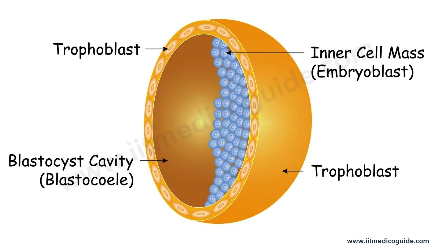
- The morula becomes a blastocyst: an outer layer, the trophoblast, and an inner cell mass. The trophoblast attaches to the endometrium; the inner cell mass becomes the embryo.
- Uterine cells divide and cover the blastocyst, embedding it in the endometrium. This is implantation, and it leads to pregnancy.
Pregnancy and embryonic development
- After implantation, finger-like chorionic villi grow from the trophoblast and interdigitate with uterine tissue to form the placenta, the structural and functional link between foetus and mother. It supplies oxygen and nutrients and removes CO2 and waste, through the umbilical cord.
- The placenta is also an endocrine tissue: it produces hCG (human chorionic gonadotropin), hPL (human placental lactogen), oestrogens and progestogens. Late in pregnancy, the ovary secretes relaxin. hCG, hPL and relaxin are produced only during pregnancy. Levels of oestrogens, progestogens, cortisol, prolactin and thyroxine also rise.
- The inner cell mass forms ectoderm and endoderm, and soon mesoderm between them; these give rise to all tissues. The inner cell mass contains stem cells, which can form all tissues and organs.
| Time | Development |
|---|---|
| End of 1st month | Heart forms; heartbeat can be heard with a stethoscope |
| End of 2nd month | Limbs and digits develop |
| End of 12 weeks (1st trimester) | Most major organ systems formed; limbs and external genitalia well developed |
| 5th month | First movements of the foetus; hair on the head appears |
| End of 24 weeks (2nd trimester) | Body covered with fine hair; eyelids separate; eyelashes form |
| End of 9 months | Foetus fully developed, ready for delivery |
Parturition and lactation
- The average human gestation period is about 9 months. Delivery by vigorous uterine contraction is parturition.
- Signals from the fully developed foetus and placenta cause mild uterine contractions: the foetal ejection reflex. This releases oxytocin from the maternal pituitary; oxytocin causes stronger contractions, which cause more oxytocin release. This positive loop expels the baby through the birth canal; the placenta follows.
- Milk production towards the end of pregnancy is lactation. The milk of the first few days, colostrum, contains several antibodies essential for the newborn's resistance. Breastfeeding is recommended for a healthy baby.
 Common traps: (1) LH acts on Leydig cells; FSH on Sertoli cells. (2) Fertilisation is in the ampulla, not the uterus. (3) Meiosis II of the oocyte completes only after sperm entry. (4) Primary oocytes are arrested in prophase I from before birth. (5) hCG, hPL and relaxin are pregnancy-only hormones. (6) The father's sperm decides the sex of the child.
NEET focus
- Duct path in males; accessory glands and seminal plasma contents.
- Parts of the oviduct and layers of the uterus; mammary gland structure.
- Spermatogenesis stages with chromosome numbers; hormonal control; sperm numbers for fertility.
- Oogenesis follicle stages; 60,000 to 80,000 follicles at puberty.
- Menstrual phases with hormones and the LH surge.
- Fertilisation, cleavage, blastocyst, implantation; placental hormones; month-wise development; parturition.
Practice questions
Androgens are secreted by:
- Sertoli cells
- Leydig cells
- Spermatogonia
- Epididymis
Show answer
The number of chromosomes in a secondary spermatocyte of a human is:
- 46
- 23
- 92
- 44
Show answer
Ovulation is induced by:
- Rise in progesterone
- LH surge
- Fall in FSH
- Oxytocin
Show answer
Fertilisation in humans normally occurs in the:
- Uterus
- Isthmus
- Ampulla of the fallopian tube
- Cervix
Show answer
Which hormone is produced only during pregnancy?
- Oestrogen
- Progesterone
- hCG
- Prolactin
Show answer
An embryo with 8 to 16 blastomeres is called a:
- Blastocyst
- Morula
- Gastrula
- Zygote
Show answer
The corpus luteum mainly secretes:
- Oestrogen
- Progesterone
- LH
- FSH