 In this chapter: types of movement; types of muscle; structure of skeletal muscle and sarcomere; actin and myosin; mechanism of contraction; red and white fibres; the human skeleton; joints; disorders of muscles and bones.
Types of movement
- Amoeboid: by pseudopodia formed by streaming of protoplasm, involving microfilaments. Seen in Amoeba and in human macrophages and leucocytes.
- Ciliary: in internal tubular organs lined by ciliated epithelium. Cilia in the trachea remove dust particles; cilia in the female reproductive tract move ova.
- Muscular: movement of limbs, jaws, tongue etc.
Locomotion is movement from one place to another. In Paramoecium, cilia help both in food movement and locomotion; in Hydra, tentacles help in capturing prey and in locomotion.
Muscle
Muscle is a specialised tissue of mesodermal origin, making up about 40 to 50% of the body weight of an adult human. Its properties are excitability, contractility, extensibility and elasticity.
| Type | Features | Location |
|---|---|---|
| Skeletal | Striated, voluntary | Attached to bones; locomotion and posture |
| Visceral (smooth) | Non-striated, involuntary | Inner walls of hollow visceral organs such as the alimentary canal and reproductive tract |
| Cardiac | Striated, involuntary, branched | Heart |
Structure of a skeletal muscle
- A muscle is made of bundles (fascicles) held together by a collagenous connective tissue layer, the fascia.
- Each fascicle contains many muscle fibres. Each fibre is lined by the sarcolemma (plasma membrane) enclosing the sarcoplasm. A fibre has many nuclei: it is a syncytium.
- The endoplasmic reticulum of muscle, the sarcoplasmic reticulum, stores calcium ions.
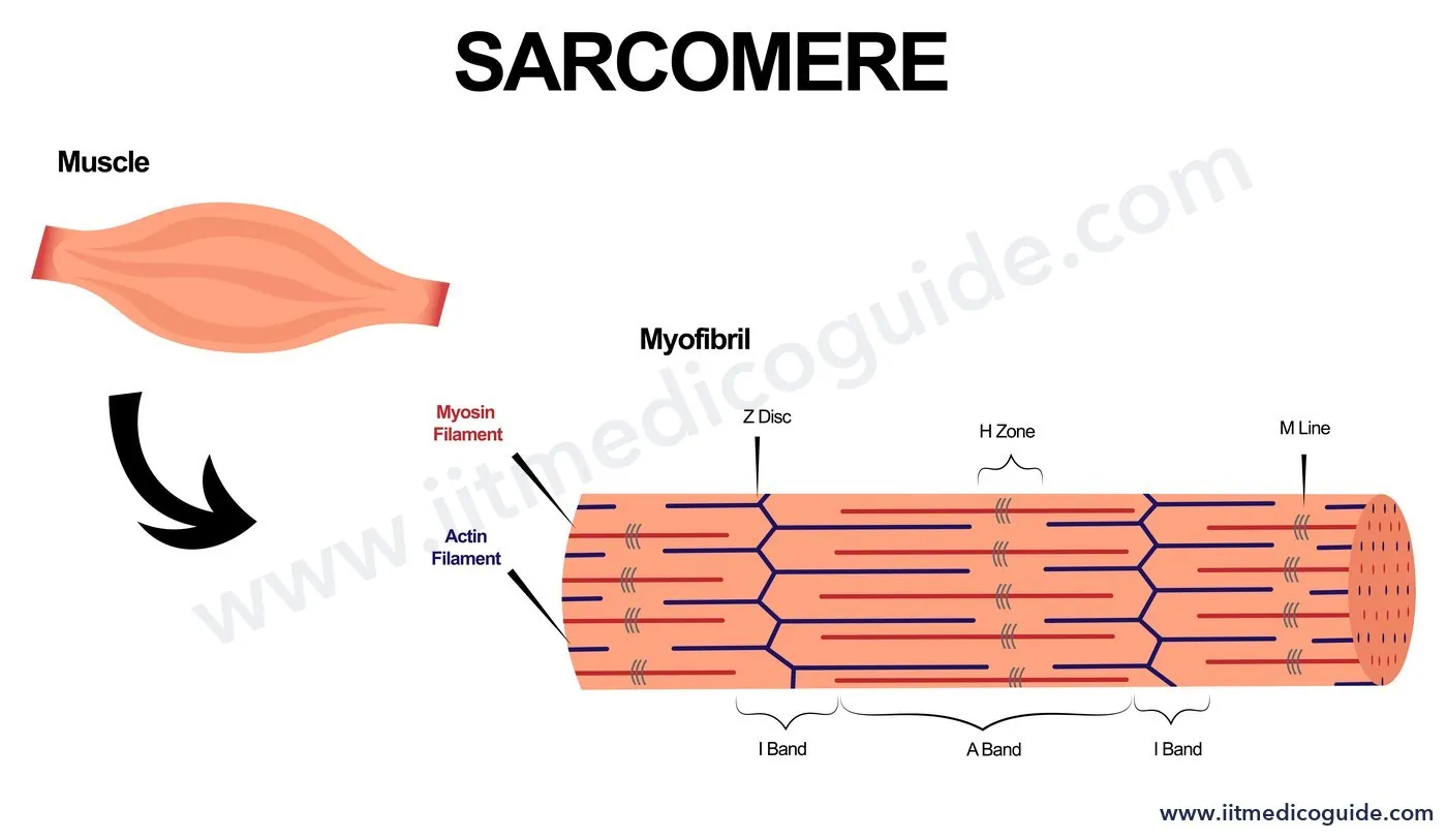
- The fibre is packed with parallel myofibrils, which show alternate dark and light bands.
The sarcomere
| Part | What it is |
|---|---|
| I band (isotropic, light) | Contains thin actin filaments only |
| A band (anisotropic, dark) | Contains thick myosin filaments (and overlapping actin) |
| Z line | Thin elastic line bisecting each I band; thin filaments are firmly attached to it |
| M line | Thin fibrous membrane in the middle of the A band holding thick filaments together |
| H zone | Central part of the thick filaments not overlapped by thin filaments (at rest) |
| Sarcomere | The portion between two successive Z lines: the functional unit of contraction |
Contractile proteins
Actin (thin) filament
Each thin filament is made of two F (filamentous) actins wound helically; each F-actin is a polymer of monomeric G (globular) actins. Two filaments of tropomyosin run along the F-actins. A complex protein, troponin, sits at regular intervals on tropomyosin. At rest, a subunit of troponin masks the active binding sites for myosin on actin.
Myosin (thick) filament
Each thick filament is a polymer of monomeric meromyosins. Each meromyosin has a globular head with a short arm (heavy meromyosin, HMM) and a tail (light meromyosin, LMM). The HMM heads project outwards at regular distances and angles as cross arms. The globular head is an active ATPase with binding sites for ATP and active sites for actin.
Mechanism of contraction: sliding filament theory
Contraction happens by thin filaments sliding over thick filaments.
- A signal from the CNS arrives through a motor neuron. A motor neuron and the muscle fibres it supplies form a motor unit. The junction between a motor neuron and the sarcolemma is the neuromuscular junction (motor end plate).
- The neural signal releases the neurotransmitter acetylcholine, generating an action potential in the sarcolemma that spreads through the fibre.
- This releases Ca2+ from the sarcoplasmic reticulum into the sarcoplasm.
- Ca2+ binds to a subunit of troponin, removing the masking of the active sites on actin.
- Using energy from ATP hydrolysis, the myosin head binds to the exposed actin sites, forming a cross bridge.
- The cross bridge pulls actin towards the centre of the A band; the Z lines are pulled inwards and the sarcomere shortens.
- Myosin releases ADP and Pi and returns to its relaxed state. A new ATP binds, the cross bridge breaks, ATP is hydrolysed, and the cycle repeats, causing further sliding.
- When Ca2+ is pumped back into the sarcoplasmic cisternae, the actin sites are masked again, the Z lines return to their original position, and the muscle relaxes.
 During contraction: the I band shortens, the H zone shortens or disappears, the A band stays the same length, and the sarcomere shortens.
Repeated activation leads to accumulation of lactic acid from anaerobic breakdown of glycogen, causing fatigue.
Red and white fibres
| Red fibres | White fibres |
|---|---|
| High myoglobin (red oxygen-storing pigment) | Less myoglobin, pale |
| Plenty of mitochondria; aerobic | Fewer mitochondria; more sarcoplasmic reticulum; depend on anaerobic metabolism |
The human skeleton
An adult human has 206 bones and a few cartilages, in the axial skeleton (80 bones) and the appendicular skeleton (126 bones).
Axial skeleton
- Skull: 8 cranial and 14 facial bones = 22. The U-shaped hyoid at the base of the buccal cavity is included with the skull. Each middle ear has three ossicles: malleus, incus and stapes. The skull joins the vertebral column through two occipital condyles (dicondylic skull).
- Vertebral column: 26 vertebrae. Each has a central neural canal for the spinal cord. The first vertebra, the atlas, articulates with the occipital condyles. Regions: cervical 7, thoracic 12, lumbar 5, sacral 1 (fused), coccygeal 1 (fused). Almost all mammals, including humans, have seven cervical vertebrae.
- Sternum: a flat bone on the ventral midline of the thorax.
- Ribs: 12 pairs, each bicephalic (two articulating surfaces at the dorsal end). Pairs 1 to 7 are true ribs (attached to the sternum by hyaline cartilage). Pairs 8, 9 and 10 are vertebrochondral (false) ribs (they join the 7th rib by hyaline cartilage). Pairs 11 and 12 are floating ribs (not connected ventrally). Thoracic vertebrae, ribs and sternum form the rib cage.
Appendicular skeleton
| Forelimb (30 bones each) | Hindlimb (30 bones each) |
|---|---|
| Humerus | Femur (thigh bone, the longest bone) |
| Radius and ulna | Tibia and fibula |
| Carpals (wrist): 8 | Tarsals (ankle): 7 |
| Metacarpals (palm): 5 | Metatarsals: 5 |
| Phalanges (digits): 14 | Phalanges: 14 |
| – | Patella (knee cap): 1 |
- Pectoral girdle: each half has a clavicle (collar bone, long, slender, with two curvatures) and a scapula (large, triangular, flat, in the dorsal thorax between the 2nd and 7th ribs). The scapula's ridge, the spine, projects as the flat acromion, with which the clavicle articulates. Below the acromion, the glenoid cavity receives the head of the humerus to form the shoulder joint.
- Pelvic girdle: two coxal bones, each formed by fusion of the ilium, ischium and pubis. At their fusion is a cavity, the acetabulum, where the femur articulates. The two halves meet ventrally at the pubic symphysis (fibrous cartilage).
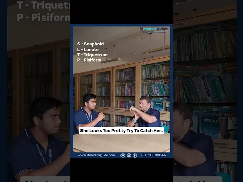
Watch a quick trick for the wrist bones from our Biology faculty:
Joints
| Type | Movement | Example |
|---|---|---|
| Fibrous | None | Sutures between flat skull bones forming the cranium |
| Cartilaginous | Limited | Between adjacent vertebrae |
| Synovial: ball and socket | Considerable (fluid-filled synovial cavity) | Humerus and pectoral girdle (shoulder) |
| Synovial: hinge | Knee | |
| Synovial: pivot | Between atlas and axis | |
| Synovial: gliding | Between carpals | |
| Synovial: saddle | Between carpal and metacarpal of the thumb |
Disorders
- Myasthenia gravis: auto-immune disorder of the neuromuscular junction causing fatigue, weakening and paralysis of skeletal muscle.
- Muscular dystrophy: progressive degeneration of skeletal muscle, mostly genetic.
- Tetany: rapid spasms (wild contractions) of muscle due to low Ca2+ in body fluid.
- Arthritis: inflammation of joints.
- Osteoporosis: age-related decrease in bone mass and higher risk of fractures; low oestrogen is a common cause.
- Gout: inflammation of joints due to accumulation of uric acid crystals.
 Common traps: (1) The A band does not shorten during contraction; the I band does. (2) Troponin masks the actin sites; Ca2+ binds troponin, not myosin. (3) Ribs 8 to 10 are vertebrochondral, not floating. (4) Tetany = low calcium; gout = uric acid. (5) Knee = hinge; atlas-axis = pivot; thumb = saddle.
NEET focus
- Sarcomere bands and what happens to each during contraction.
- Roles of troponin, tropomyosin, HMM and LMM; the contraction sequence.
- Bone counts: 206, 80, 126, 22, 26, 12 pairs of ribs, 30 per limb; vertebral formula.
- True, false and floating ribs; parts of the girdles; joint types with examples; disorders.
Practice questions
During muscle contraction, which band keeps its length?
- I band
- A band
- H zone
- Sarcomere
Show answer
Calcium ions released during muscle excitation bind to:
- Myosin head
- Tropomyosin
- Troponin
- Actin
Show answer
The ATPase activity is present in the:
- Tail of myosin (LMM)
- Head of myosin (HMM)
- Troponin
- G-actin
Show answer
Ribs that are not connected ventrally are:
- Pairs 1 to 7
- Pairs 8 to 10
- Pairs 11 and 12
- Pair 7 only
Show answer
The joint between atlas and axis is a:
- Hinge joint
- Pivot joint
- Saddle joint
- Gliding joint
Show answer
Tetany is caused by:
- Accumulation of uric acid
- Low Ca2+ in body fluid
- Auto-immune attack on neuromuscular junction
- Low oestrogen
Show answer
The number of bones in the human axial skeleton is:
- 126
- 80
- 206
- 22