

 In this chapter: ammonotelism, ureotelism, uricotelism; excretory structures in animals; human kidney and nephron; filtration, reabsorption, secretion; functions of each tubule segment; counter-current mechanism; regulation (ADH, renin-angiotensin, ANF); micturition; other excretory organs; disorders and dialysis.
Modes of excretion
| Mode | Main waste | Why | Examples |
|---|---|---|---|
| Ammonotelism | Ammonia (most toxic, needs much water) | Readily soluble; diffuses out through body surface or gills as ammonium ions | Many bony fishes, aquatic amphibians, aquatic insects |
| Ureotelism | Urea (made in the liver from ammonia) | Less toxic, conserves water. Some urea is retained in the kidney matrix of some animals to maintain osmolarity | Mammals, many terrestrial amphibians, marine fishes |
| Uricotelism | Uric acid (pellet or paste, minimum water loss) | Least toxic, least water needed | Reptiles, birds, land snails, insects |
Excretory structures in animals
- Protonephridia (flame cells): Platyhelminthes (e.g. Planaria), rotifers, some annelids and the cephalochordate Amphioxus. Mainly for osmoregulation (ionic and fluid balance).
- Nephridia: earthworms and other annelids.
- Malpighian tubules: most insects including cockroaches.
- Antennal (green) glands: crustaceans like prawns.
The human excretory system
It consists of a pair of kidneys, a pair of ureters, a urinary bladder and a urethra.
- Kidneys are reddish-brown, bean-shaped, lying between the levels of the last thoracic and third lumbar vertebra, close to the dorsal inner wall of the abdominal cavity. Each is 10 to 12 cm long, 5 to 7 cm wide, 2 to 3 cm thick, weighing 120 to 170 g.
- The inner concave side has a notch, the hilum, through which the ureter, blood vessels and nerves pass. Inside is the funnel-shaped renal pelvis with projections called calyces.
- Inside, the kidney has an outer cortex and an inner medulla. The medulla has conical medullary pyramids projecting into the calyces. The cortex extends between the pyramids as renal columns (columns of Bertini).
- Each kidney has about one million nephrons, the functional units.
The nephron
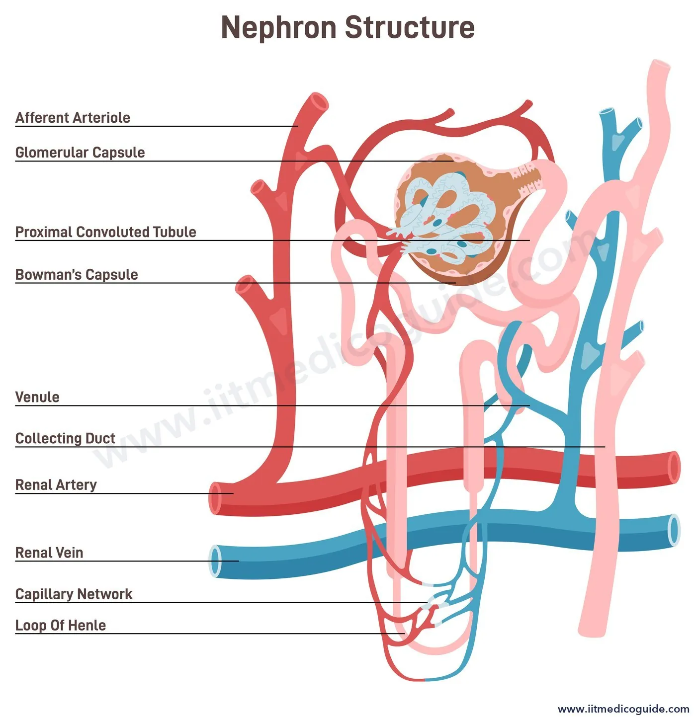
- Glomerulus: a tuft of capillaries formed by the afferent arteriole (a fine branch of the renal artery); blood leaves through the efferent arteriole.
- Bowman's capsule: a double-walled cup around the glomerulus. Glomerulus + Bowman's capsule = Malpighian body (renal corpuscle).
- The tubule continues as the highly coiled PCT (proximal convoluted tubule), the hairpin-shaped Henle's loop (descending and ascending limbs) and the highly coiled DCT (distal convoluted tubule). DCTs of many nephrons open into a straight collecting duct; collecting ducts open into the renal pelvis through the pyramids.
- The Malpighian corpuscle, PCT and DCT lie in the cortex; the loop of Henle dips into the medulla.
- Cortical nephrons (the majority) have a short loop that extends only a little into the medulla. Juxtamedullary nephrons have very long loops running deep into the medulla.
- The efferent arteriole forms the peritubular capillaries around the tubule. A minute vessel of this network runs parallel to Henle's loop as a U-shaped vasa recta, which is absent or highly reduced in cortical nephrons.
Urine formation
1. Glomerular filtration
- Kidneys filter about 1100 to 1200 mL of blood per minute, roughly one-fifth of the blood pumped by each ventricle.
- Glomerular capillary blood pressure drives filtration through three layers: the endothelium of glomerular capillaries, the epithelium of Bowman's capsule and the basement membrane between them. The capsule's epithelial cells, podocytes, leave minute spaces called filtration slits (slit pores).
- Almost everything in plasma except proteins passes through: this is ultrafiltration.
- GFR (glomerular filtration rate) in a healthy person is about 125 mL per minute, i.e. 180 litres per day.
- The juxtaglomerular apparatus (JGA) is a sensitive region formed where the DCT touches the afferent arteriole. A fall in GFR makes JG cells release renin, which restores glomerular blood flow and GFR.
2. Reabsorption
Of 180 L of filtrate per day, only about 1.5 L becomes urine, so nearly 99% is reabsorbed. Glucose, amino acids and Na+ are reabsorbed actively; nitrogenous wastes passively; water passively in the initial segments.
3. Tubular secretion
Tubular cells secrete H+, K+ and ammonia into the filtrate, helping maintain ionic and acid-base balance.
What each part does
| Segment | Function |
|---|---|
| PCT | Simple cuboidal brush border epithelium. Reabsorbs nearly all essential nutrients and 70 to 80% of electrolytes and water. Maintains pH by secreting H+, ammonia and K+ and absorbing HCO3− |
| Descending limb of Henle's loop | Permeable to water, almost impermeable to electrolytes: filtrate becomes concentrated |
| Ascending limb of Henle's loop | Impermeable to water, transports electrolytes actively or passively: filtrate becomes dilute. Helps maintain high osmolarity of the medullary interstitium |
| DCT | Conditional reabsorption of Na+ and water; reabsorbs HCO3−; secretes H+, K+ and NH3 to maintain pH and Na-K balance |
| Collecting duct | Reabsorbs large amounts of water to make concentrated urine; lets small amounts of urea into the medullary interstitium to keep up osmolarity; secretes H+ and K+ |
Counter-current mechanism
Filtrate flows in opposite directions in the two limbs of Henle's loop, and blood flows in opposite directions in the two limbs of the vasa recta. Both are counter currents. Their closeness helps build an increasing osmolarity towards the inner medulla: from about 300 mOsmol L−1 in the cortex to about 1200 mOsmol L−1 in the inner medulla.
- This gradient is caused mainly by NaCl and urea.
- NaCl transported out of the ascending limb of Henle's loop is exchanged with the descending limb of the vasa recta, and returned to the interstitium by the ascending portion of the vasa recta.
- Small amounts of urea enter the thin segment of the ascending limb and are returned to the interstitium by the collecting tubule.
- The gradient lets water leave the collecting duct easily, so mammals can make urine nearly four times as concentrated as the initial filtrate.
Regulation of kidney function
- ADH (vasopressin): osmoreceptors in the hypothalamus detect loss of body fluid; the hypothalamus causes release of ADH from the neurohypophysis. ADH increases water reabsorption from the latter parts of the tubule, preventing diuresis. More body fluid switches the receptors off and suppresses ADH. ADH also constricts blood vessels, raising blood pressure and GFR.
- Renin-angiotensin mechanism: a fall in glomerular blood flow, blood pressure or GFR activates JG cells to release renin. Renin converts angiotensinogen to angiotensin I, which becomes angiotensin II, a powerful vasoconstrictor that raises glomerular blood pressure and GFR. Angiotensin II also makes the adrenal cortex release aldosterone, which increases reabsorption of Na+ and water from the distal tubule, raising blood pressure and GFR.
- ANF (atrial natriuretic factor): increased blood flow to the atria of the heart causes ANF release, which dilates blood vessels and lowers blood pressure. ANF acts as a check on the renin-angiotensin mechanism.
Micturition
Urine is stored in the bladder until a voluntary signal from the CNS. As the bladder fills and stretches, stretch receptors send signals to the CNS, which sends motor messages to contract the bladder's smooth muscles and relax the urethral sphincter. This is the micturition reflex.
An adult excretes 1 to 1.5 litres of urine a day. It is light yellow and slightly acidic (pH about 6.0), with a characteristic odour, and on average 25 to 30 g of urea is excreted a day. Glucose (glycosuria) or ketone bodies (ketonuria) in urine suggest diabetes mellitus.
Other organs that help in excretion
- Lungs: remove about 200 mL of CO2 per minute and significant water.
- Liver: the largest gland; bile carries bilirubin, biliverdin, cholesterol, degraded steroid hormones, vitamins and drugs, which leave with digestive waste.
- Skin: sweat (water with NaCl, small amounts of urea, lactic acid etc., mainly for cooling) and sebum from sebaceous glands (sterols, hydrocarbons, waxes: a protective oily covering).
- Saliva: small amounts of nitrogenous waste.
Disorders
- Uraemia: accumulation of urea in blood; can lead to kidney failure. Treated by haemodialysis: blood from an artery, with an anticoagulant like heparin added, is pumped through a coiled cellophane tube in dialysing fluid that has the same composition as plasma but no nitrogenous wastes. Wastes diffuse out down their concentration gradient; the cleaned blood, with anti-heparin added, returns through a vein.
- Kidney transplantation: the ultimate treatment for acute renal failure; a kidney from a close relative is preferred to reduce rejection.
- Renal calculi: stones of crystallised salts (oxalates etc.) in the kidney.
- Glomerulonephritis: inflammation of the glomeruli.
 Common traps: (1) Descending limb is permeable to water; ascending limb is not. (2) Vasa recta is reduced or absent in cortical nephrons. (3) ANF opposes the renin-angiotensin system. (4) Marine fishes are ureotelic; many bony fishes (freshwater) are ammonotelic. (5) GFR is 125 mL/min, but renal blood flow is 1100 to 1200 mL/min.
NEET focus
- Modes of excretion with examples; excretory organs of invertebrates.
- Kidney position and dimensions; nephron parts and their location; cortical vs juxtamedullary.
- GFR, 99% reabsorption, podocytes; segment-wise functions.
- Counter-current numbers (300 to 1200 mOsmol); ADH, renin-angiotensin-aldosterone, ANF.
- Urine composition; dialysis steps.
Practice questions
The normal GFR in a healthy person is about:
- 1200 mL/min
- 125 mL/min
- 180 mL/min
- 1.5 L/min
Show answer
Which part of the nephron is impermeable to water?
- PCT
- Descending limb of Henle's loop
- Ascending limb of Henle's loop
- Collecting duct (with ADH)
Show answer
About 70 to 80% of electrolytes and water are reabsorbed in the:
- PCT
- DCT
- Henle's loop
- Collecting duct
Show answer
Which hormone acts as a check on the renin-angiotensin mechanism?
- Aldosterone
- ADH
- ANF
- Angiotensin II
Show answer
Uricotelism is found in:
- Bony fishes
- Mammals
- Birds and reptiles
- Aquatic amphibians
Show answer
Filtration slits are formed by:
- Endothelial cells
- Podocytes
- JG cells
- Brush border cells
Show answer
The osmolarity of the inner medullary interstitium reaches about:
- 300 mOsmol L−1
- 600 mOsmol L−1
- 1200 mOsmol L−1
- 2400 mOsmol L−1